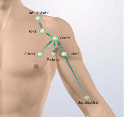
axillary lymph node level
PPT - IMAGING OF THE AXILLA PowerPoint Presentation, free download - ID. 16 Pictures about PPT - IMAGING OF THE AXILLA PowerPoint Presentation, free download - ID : PPT - IMAGING OF THE AXILLA PowerPoint Presentation - ID:6722507, Breast Procedures and also Utility of Level III Axillary Lymph Node Dissection in Melanoma.
PPT - IMAGING OF THE AXILLA PowerPoint Presentation, Free Download - ID
 www.slideserve.com
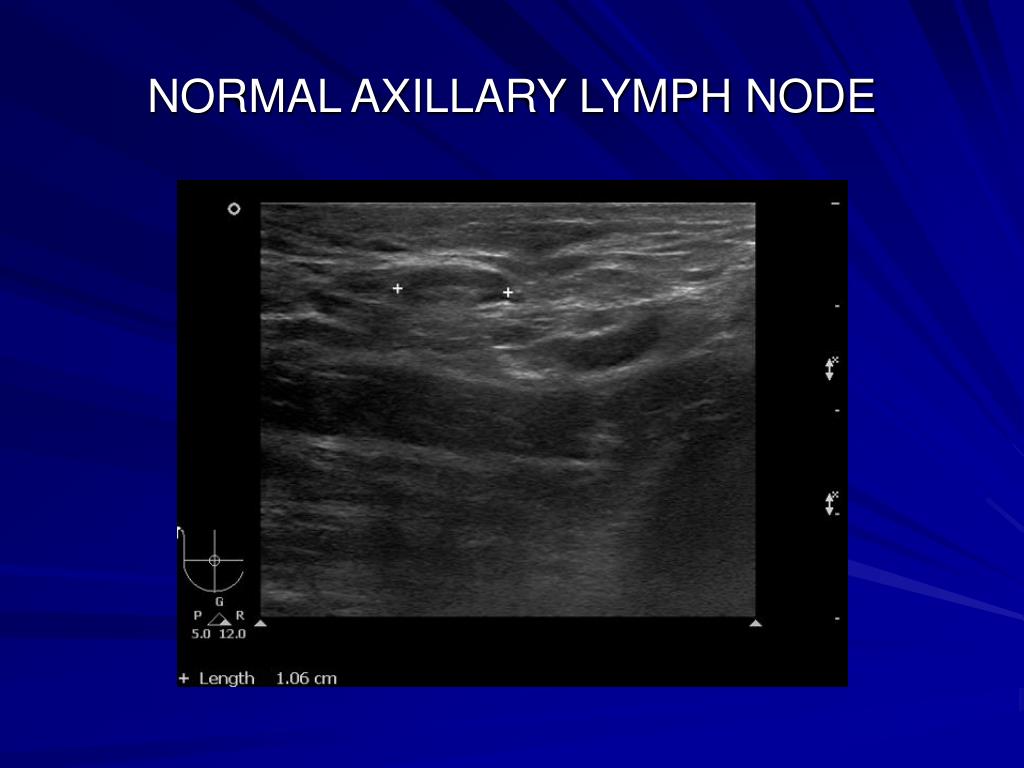
normal axillary lymph node axilla
CHAPTER 39 Transverse Myocutaneous Gracilis With Vascularized Lymph
 plasticsurgerykey.com
lymph node transfer transverse myocutaneous gracilis flap vascularized chapter fig surgery positioned breast chest finished
Normal Lymph Node: On Ultrasound, Lymph Nodes Typically Are Smooth
 www.researchgate.net
ultrasound lymph node nodes cortex typically hilum lobulated gently hypoechoic fig12 priscilla
Reactive Axillary Lymph Node | Radiology Case | Radiopaedia.org
 radiopaedia.org
radiopaedia
Breast Procedures
 www.medscape.com
lymph axillary dissection axilla shown
PPT - IMAGING OF THE AXILLA PowerPoint Presentation - ID:6722507
 www.slideserve.com
lymph axilla axillary
Lymph Node Size Chart - Best Picture Of Chart Anyimage.Org
 www.rechargecolorado.org
lymph axillary nodes patients micrometastases immunohistochemical
Utility Of Level III Axillary Lymph Node Dissection In Melanoma
 www.journalacs.org
s250 oncology
Pin On ANATOMY
 www.pinterest.com
lymph node sentinel nodes neck head anatomy lymphatic melanoma biopsy surgical glands levels metastasis approach intechopen 5a vessels
Reactive Axillary Lymph Node | Image | Radiopaedia.org
 radiopaedia.org
lymph reactive axillary radiopaedia
Pectoral Girdle And Upper Limb: Overview And Surface Anatomy
 basicmedicalkey.com
upper limb lymph nodes pectoral anatomy lymphatic left drainage surface girdle fig key
ABC Of Breast Diseases: Management Of Regional Nodes In Breast Cancer
 www.bmj.com
nodes axillary bmj
Axillary Lymph Nodes Examination - YouTube
 www.youtube.com
axillary lymph
Lymph Nodes Axial 6
 www.netterimages.com
lymph axial
Imaging Findings Of Variable Axillary Mass And Axillary Lymphadenopathy
 www.umbjournal.org
axillary lymphadenopathy
Intercostobrachial Nerves As A Novel Anatomic Landmark For Dividing The
 www.hindawi.com
axillary dissection lymph node space intercostobrachial nerves landmark dividing anatomic novel figure
Ultrasound lymph node nodes cortex typically hilum lobulated gently hypoechoic fig12 priscilla. Axillary lymph nodes examination. Reactive axillary lymph node