foot skeleton xray
Foot Problems and Foot Pain. 16 Pictures about Foot Problems and Foot Pain : Foot Bones X Ray / Cureus Chondromyxoid Fibroma Of Distal Phalanx Of, Tarsal Bones; Ankle Bones; Cuboid Bone; Cuneiform Bones; Navicular Bone and also X Ray Foot Anatomy - Anatomy Drawing Diagram.
Foot Problems And Foot Pain
 footpainrx.blogspot.com
foot pain problems extra bones
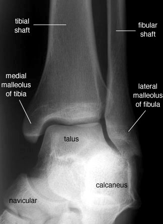
Radiographic Anatomy Of The Skeleton: Ankle -- Mortise View, Labelled
 uwmsk.org
mortise ankle radiology anatomy labelled radiographic foot labeled lateral oblique medical talus ortopedia anatomical unlabelled version
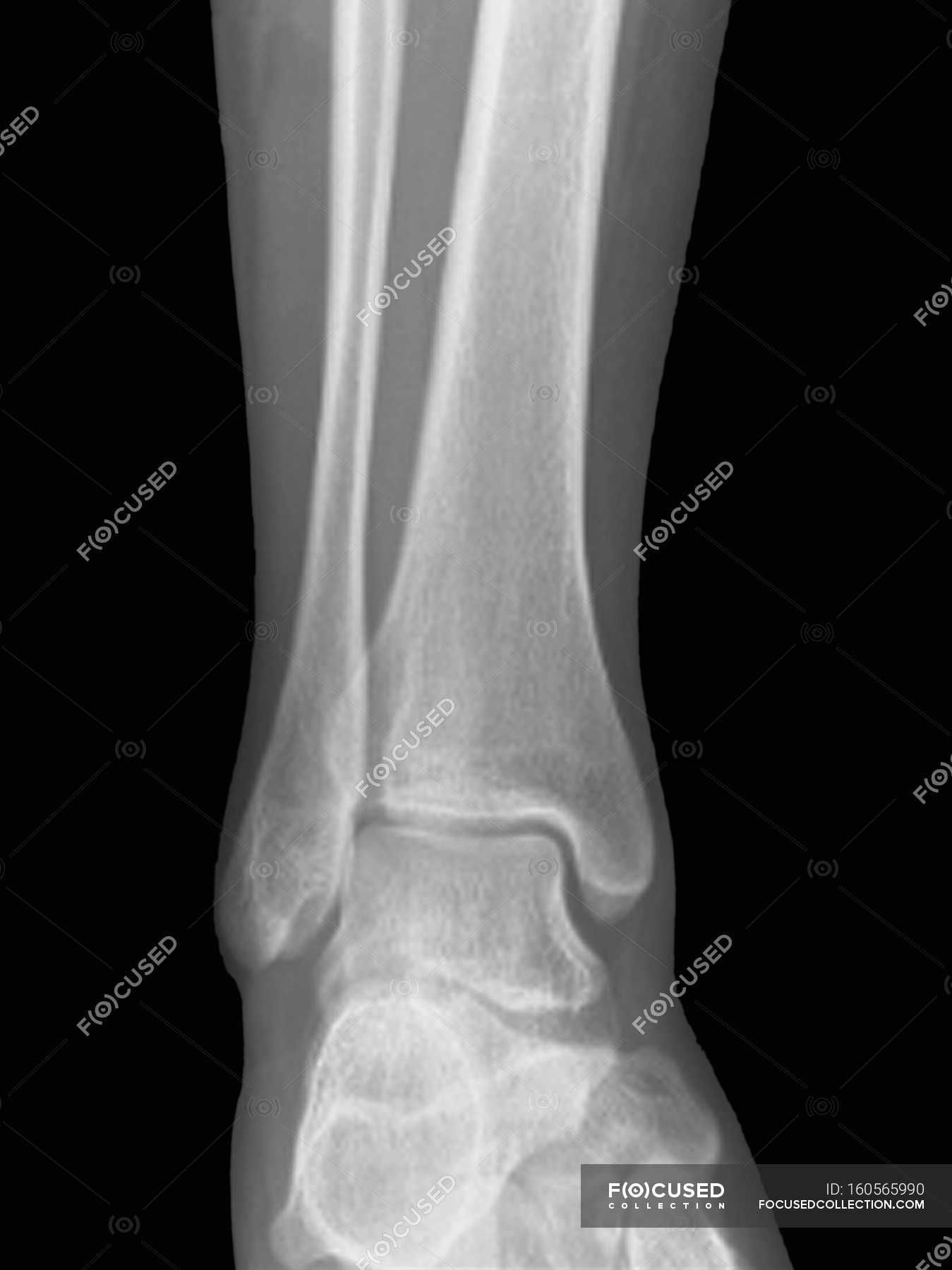
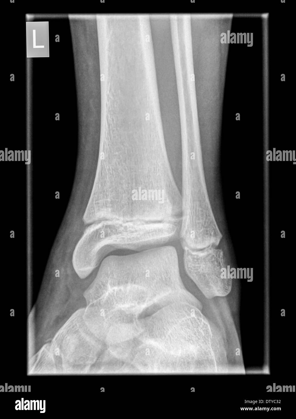
Normal Ankle Joint, Frontal X-ray. — Front View, Human Body - Stock
 focusedcollection.com
ankle frontal
Bones Of The Foot Xray
 www.nbu.bg
xray labeled
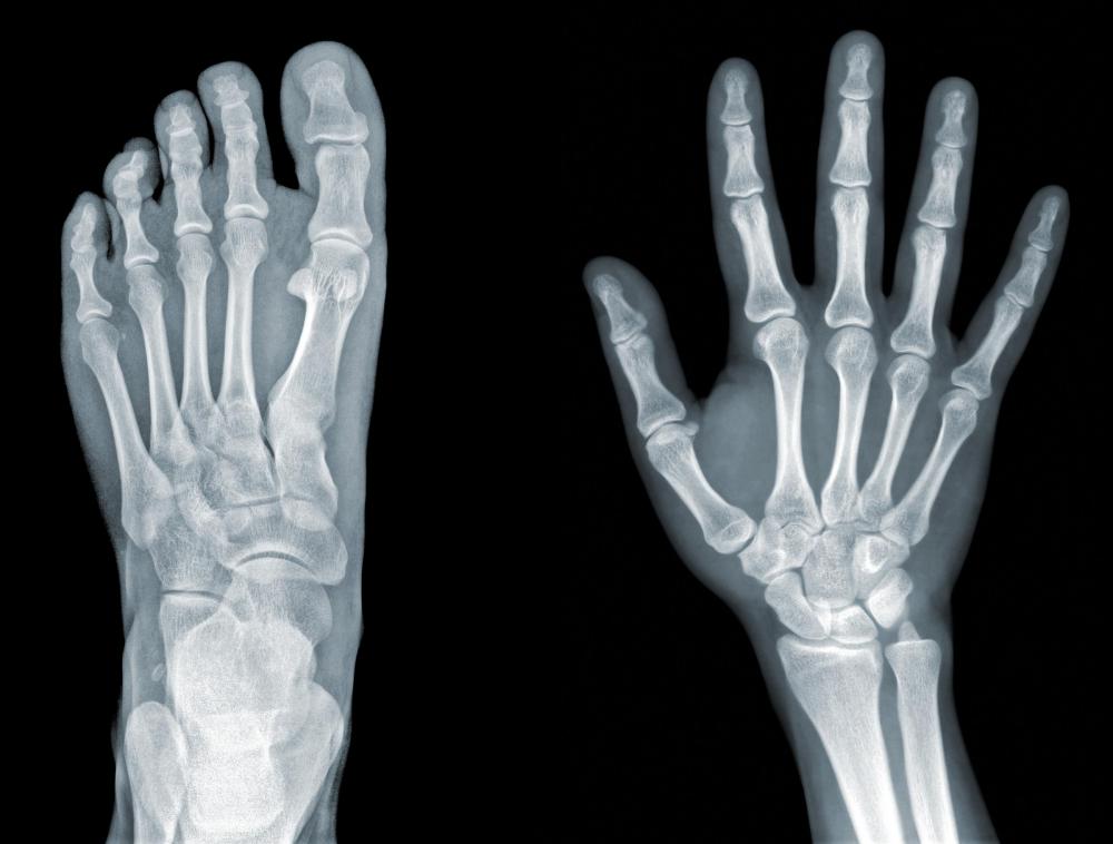
What Is An Accessory Bone? (with Pictures)
 www.wisegeek.com
bone accessory bones extra hand ray foot body
Broken Right Foot Ankle Xray. Stock Image - Image Of Doctor, Broken
 www.dreamstime.com
foot xray right ankle broken side premium
"Top View Of Humans Feet Bones Under X-ray" Stock Photo And Royalty
 www.fotolia.com
feet
Coloured X-ray Of The Bones In The Foot - Stock Image - P116/0408
 www.sciencephoto.com
coloured
Brian's Running Adventures: January 2013
 briansrunningadventures.blogspot.com
foot bones january feet wednesday each there
Normal Bone Of Human's Foot Royalty Free Stock Photography - Image
 www.dreamstime.com
foot human bone normal royalty ray film orthopedic surgery dreamstime
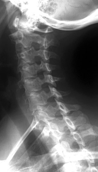
Radiographic Anatomy Of The Skeleton: Cervical Spine -- Left Anterior
 uwmsk.org
spine cervical ray oblique labelled radiology anatomy left radiographic anterior neck diagram skeleton normal imaging vertebrae uwmsk tech xray schools
Tarsal Bones; Ankle Bones; Cuboid Bone; Cuneiform Bones; Navicular Bone
 www.lookfordiagnosis.com
os foot bone navicular tibiale accessory bones externum tarsal cuboid cuneiform rays fuss roe ankle ray file medial peroneum commons
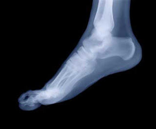
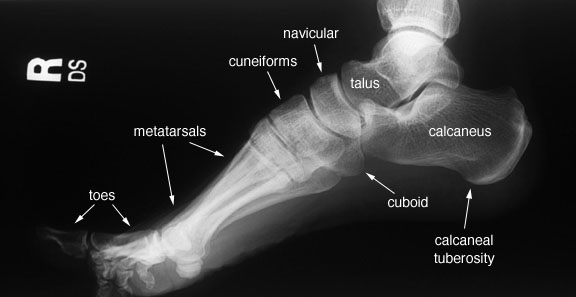
X Ray Foot Anatomy - Anatomy Drawing Diagram
 sen842cova.blogspot.com
lateral foot ray anatomy radiographic radiology labelled skeleton bones uwmsk radiography medical enregistrée depuis imaging
Bare Bones – WOW! Children's Museum
 wowchildrensmuseum.org
bones children foot xray bare wow museum
Healthy X-ray Of An Ankle 12 Year Old Male Front View Stock Photo
 www.alamy.com
Foot Bones X Ray / Cureus Chondromyxoid Fibroma Of Distal Phalanx Of
 lucindaj-sand.blogspot.com
sand
Foot bones january feet wednesday each there. Lateral foot ray anatomy radiographic radiology labelled skeleton bones uwmsk radiography medical enregistrée depuis imaging. Mortise ankle radiology anatomy labelled radiographic foot labeled lateral oblique medical talus ortopedia anatomical unlabelled version